
Google researchers, in collaboration with Harvard University, have reconstructed a tiny piece of the human brain at an unprecedented level of detail, revealing never-before-seen structures and paving the way for groundbreaking discoveries in neuroscience.
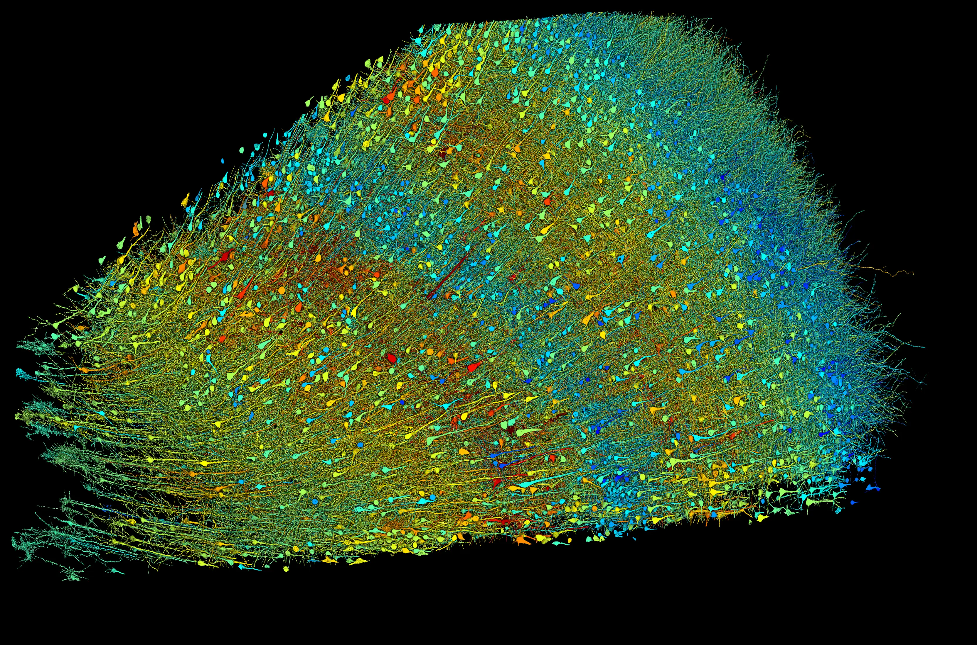 Researchers built a 3D image of nearly every neuron and their connections within a small piece of human brain tissue. The above image shows excitatory neurons and the banner image shows inhibitory neurons. These versions are shaded according to the size of the neurons’ cell bodies (central core), which range from 15–30 micrometers across. The sample is approximately 3 mm wide. Credit: Google Research & Lichtman Lab (Harvard University). Renderings by D. Berger (Harvard University).
Understanding the human brain, with its intricate network of billions of neurons and trillions of connections, has long been a scientific quest. While we’ve made significant strides in understanding broad brain functions and regions, the precise map of how individual brain cells connect and communicate remains largely unexplored. This is where the field of connectomics comes in, aiming to create comprehensive maps of neural connections to unlock the secrets of brain function and dysfunction.
Marking a decade of research in connectomics, the Google AI team, in partnership with Harvard University, has achieved a remarkable feat: reconstructing a small section of the human brain at nanoscale resolution. This groundbreaking work, published in the journal Science, provides an unprecedented glimpse into the intricate architecture of the human brain, revealing previously unknown structures and offering new avenues for understanding brain function, development, and disease.
 The study focused on a cubic millimeter of brain tissue, about the size of a grain of rice, extracted from the temporal lobe of a patient undergoing epilepsy surgery. Using advanced imaging techniques and powerful AI algorithms, the researchers meticulously reconstructed the 3D structure of nearly 57,000 cells, including neurons, glial cells, and blood vessels. This detailed reconstruction unveiled the intricate network of connections between these cells, mapping over 150 million synapses.
One of the most striking discoveries was the presence of exceptionally strong synaptic connections between certain pairs of neurons. These rare connections, involving multiple synapses between two cells, suggest a mechanism for particularly fast or crucial neural communication, potentially playing a role in rapid information processing or the formation of strong memories.
Further intriguing observations included a curious mirror symmetry in the arrangement of certain neurons in the deepest layer of the cortex, hinting at an underlying functional organization yet to be deciphered. Additionally, the researchers identified enigmatic “axon whorls,” where axons, the long projections of neurons, form intricate knots, the purpose of which remains a mystery.
The researchers emphasize that these findings are just the tip of the iceberg, with the vast dataset holding the potential for numerous further discoveries. To facilitate exploration and collaboration, the team has developed open-source tools like Neuroglancer and CAVE, allowing researchers worldwide to delve into this intricate brain map and contribute to its refinement.
Looking ahead, the team is working on scaling up connectomics to larger brain regions and exploring its applications in understanding neurological diseases such as Alzheimer’s. By unraveling the complex wiring of the human brain, connectomics promises to revolutionize our understanding of the brain in health and disease, paving the way for novel therapies and treatments.
Link to the original article at Google Research blog.
Link to the original research article in Science magazine.


Responses (0 )